


Rare Diseases in Numbers
Rare diseases encompass a broad and heterogeneous group of conditions that…
Continue reading
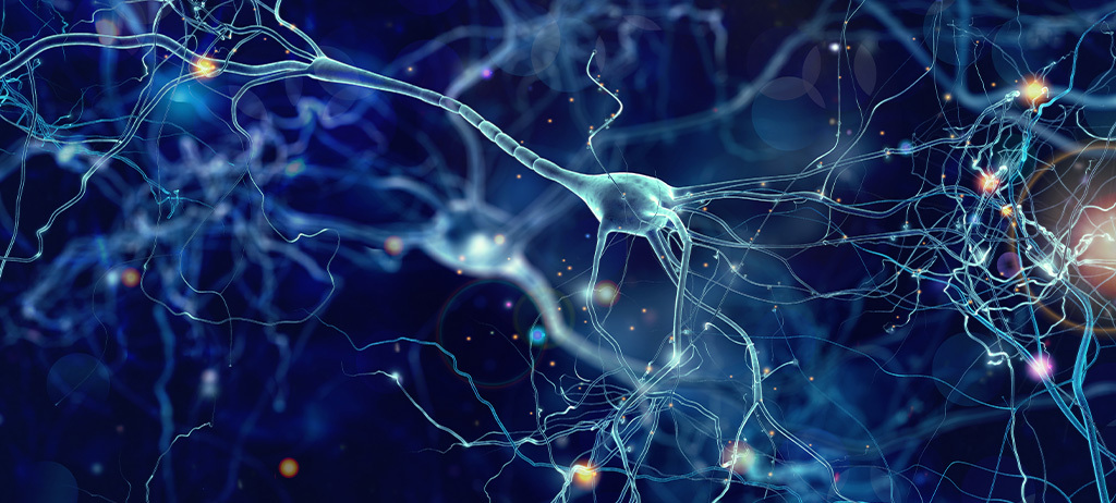
Early diagnosis and monitoring of neurological diseases represent some of the greatest challenges in modern medicine. With advances in neuroscience and laboratory technologies, it has become possible to investigate specific biomarkers that directly reflect neuronal integrity and damage. Among these biomarkers, neurofilaments—especially neurofilament light chain (NfL)—have stood out for their sensitivity and applicability in various clinical contexts, such as multiple sclerosis (MS), amyotrophic lateral sclerosis (ALS), and Alzheimer’s disease (AD).
Neurofilaments are essential structural proteins of the neuronal cytoskeleton, particularly abundant in axons. When damage to the central or peripheral nervous system occurs, these proteins are released into the cerebrospinal fluid (CSF) and the bloodstream, making their detection possible through laboratory tests.
In this article, we provide a comprehensive analysis of the role of neurofilaments as promising biomarkers for early diagnosis, prognosis, and monitoring of various neurological diseases. We also discuss their structural characteristics, the pathophysiology behind their release into bodily fluids, and the technological advances that have enabled their clinical use. We explore the main scientific evidence supporting their utility in conditions such as multiple sclerosis, amyotrophic lateral sclerosis, Alzheimer’s disease, Parkinson’s disease, among others. Enjoy your reading!
Neurofilaments (NFs) are structural proteins that form part of the cytoskeleton of neurons, particularly abundant in axons (1). They belong to the intermediate filament family and are essential for maintaining axonal caliber and the integrity of axoplasmic transport.
Neurofilaments are composed of three major subunits, classified according to their size and function (1, 2):
Because they are neuron-specific proteins and released into bodily fluids after axonal injury, neurofilaments have emerged as promising biomarkers for neurological diseases.
Neurofilaments are essential structural components of the neuronal cytoskeleton and play a fundamental role in maintaining the integrity and functionality of the axon—the long extension of the nerve cell responsible for conducting electrical impulses.
The primary function of neurofilaments is to provide mechanical support by regulating axonal diameter, which directly influences nerve conduction velocity. In addition, neurofilaments are actively involved in axonal transport, assisting in the movement of organelles and proteins along the axon—an essential process for the survival and proper function of neurons (3).
In the event of axonal damage, as seen in neurodegenerative processes or trauma, neurofilaments are released into the extracellular space (4) and can be detected in the cerebrospinal fluid (CSF) and, to a lesser extent, in the peripheral blood. This release allows for the use of neurofilaments—especially the light chain (NfL)—as sensitive biomarkers of neuronal injury.
Neurofilament light chain (NfL) is the most abundant and soluble subunit of neurofilaments and is the first to be detected in circulation in response to neuronal damage. For this reason, it is considered a highly sensitive biomarker for various neurological disorders.
NfL has the lowest molecular weight among the neurofilament subunits and plays a crucial role in axonal structural stabilization and axon growth promotion (5, 6).
In healthy individuals, NfL is detectable at low levels in both blood and CSF, with a progressive increase associated with aging. In the presence of neuronal injury or degeneration, its release into bodily fluids becomes more pronounced, likely due to loss of cellular membrane integrity (6).
Studies have shown that elevated NfL levels in blood or CSF are associated with the severity and progression of neurodegenerative diseases, inflammatory, and traumatic conditions such as Alzheimer’s disease (AD) (7). In addition to its stability, the ability to measure it in plasma facilitates its use in large-scale clinical settings. Animal models also support its utility in assessing the severity of neuronal apoptosis (8, 9).
The primary purpose of neurofilament assessment is to detect and monitor axonal injury, making it useful for differential diagnosis and for monitoring treatment response in various neurological conditions.
The test can be used in patients with early neurological symptoms as well as in advanced cases to evaluate disease activity or progression. It also holds prognostic value, helping to predict the rate of progression in some neurological diseases.
Several studies have reported the potential value of CSF and blood analysis for quantifying NfL as a biomarker in various diseases characterized by axonal loss, including stroke, small vessel disease, HIV infection, traumatic brain injury, amyotrophic lateral sclerosis (ALS), Alzheimer’s disease, Huntington’s disease, acute spinal cord injury, neuromyelitis optica, and multiple sclerosis (MS) (10, 11).
Neurofilaments, especially the light chain subunit (NfL), have emerged as promising biomarkers in various neurodegenerative diseases (12–15).
As previously discussed, following axonal injury, these proteins are released into the bloodstream and cerebrospinal fluid (CSF), serving as indicators of neuronal damage—but they do not directly identify the underlying cause.
Nonetheless, they hold clinical value, as they can help differentiate neurodegenerative diseases from functional disorders, while also providing important prognostic insights. Serial testing also allows clinicians to monitor treatment efficacy or detect early relapses, as in the case of multiple sclerosis (MS).
Despite their potential, the clinical interpretation of NfL requires caution, as baseline levels can be influenced by age, body mass index, and medication use.
Furthermore, because NfL can be reliably measured in blood, the test is minimally invasive, making it suitable for repeated and longitudinal assessments over time to support clinical monitoring.

The investigation of neurofilaments, particularly NfL, has proven useful in various neurological conditions characterized by axonal injury or degeneration. Key conditions include:
In MS, a chronic inflammatory disease of the central nervous system, NfL has been established as a sensitive marker of axonal damage, reflecting both neurodegeneration and inflammation.
NfL is primarily released during inflammatory relapses or progressive phases of the disease, as a result of axonal damage caused by demyelination and chronic inflammation.
Studies show that serum NfL levels correlate with disease activity, brain atrophy, and MRI findings, such as lesion number and volume. Additionally, elevated NfL levels have prognostic value, predicting greater disability progression, especially in relapsing-remitting MS (RRMS) (16).
Quantifying NfL in peripheral blood has become a viable tool for longitudinal MS monitoring, enabling assessment of treatment response and detection of subclinical activity.
Due to its high sensitivity and specificity, NfL stands out as a multidimensional biomarker, useful for diagnosis, monitoring, and prognosis across different MS stages and subtypes (16).
Moreover, persistent NfL elevation is associated with a higher risk of progression to secondary progressive MS (SPMS) (10, 17), and ongoing monitoring supports evaluation of immunomodulatory therapy effectiveness.
Neurofilaments—especially NfL—are highly sensitive and specific biomarkers for ALS, measurable in both CSF and peripheral blood.
NfL levels are significantly elevated in ALS patients compared to healthy individuals and those with other neuromuscular disorders, reinforcing its value for differential diagnosis and early clinical confirmation, particularly in early disease stages when clinical signs may be subtle (18).
Beyond diagnostics, NfL levels are prognostic in ALS. Their concentration correlates with disease progression speed and survival time—higher levels predict a more aggressive course, while lower levels indicate slower disease progression.
This relationship supports risk stratification, individualized clinical monitoring, and therapeutic response evaluation in clinical trials. Due to its plasma stability and ease of measurement, NfL is an ideal biomarker for both clinical practice and research (18).
ALS is a rapidly progressive neurodegenerative disease affecting upper and lower motor neurons. NfL levels are elevated early in the disease and remain high throughout progression, aiding in differentiation from other motor neuron disorders and contributing to prognosis and therapeutic monitoring in both clinical and research settings.
NfL has shown great promise as a biomarker for early detection and disease progression monitoring in both AD and mild cognitive impairment (MCI). Multiple studies demonstrate significantly higher NfL levels in AD and MCI patients than in healthy controls, with the highest levels in AD. NfL is negatively correlated with Mini-Mental State Examination (MMSE) scores, suggesting an association with cognitive decline and shorter survival in dementia patients (19, 20).
Although not specific to AD, NfL reflects ongoing neuroaxonal degeneration and can be detected in preclinical stages, making it valuable for differential diagnosis, risk stratification, and evaluation of disease-modifying therapies (21).
Its plasma levels increase gradually and correlate with brain atrophy observed on neuroimaging. The main advantage of NfL is its potential for early screening, preventive medicine, and large-scale population studies (19, 21).
In PD, plasma NfL has emerged as a biomarker of neuroaxonal damage with potential clinical utility, particularly for predicting motor and cognitive progression. While early disease stages may show only slight NfL elevations, longitudinal studies reveal that higher levels are associated with greater motor deterioration—especially in the postural instability and gait difficulty (PIGD) subtype—and cognitive decline.
Patients with higher NfL levels are more likely to progress to advanced stages, require mobility aids, or become institutionalized (22).
NfL also helps in differentiating idiopathic PD from atypical parkinsonian syndromes (APS) such as multiple system atrophy and progressive supranuclear palsy, which show significantly higher NfL levels—even in early stages—supporting early clinical differentiation. Although study results vary, age-adjusted and intra-individual longitudinal evaluations appear to improve the sensitivity of detecting meaningful disease progression (22).
Other related conditions include traumatic brain injury, frontotemporal dementia, Huntington’s disease, atypical parkinsonism, autoimmune encephalopathies, and central nervous system infections.
The NfL test measures the concentration of this protein in body fluids, typically in blood (plasma or serum) or cerebrospinal fluid (CSF). NfL is a subunit of neurofilaments—key components of the neuronal cytoskeleton—released into extracellular fluid following axonal damage.
Initially, NfL quantification was restricted to CSF, limiting its use. The development of ELISA assays for peripheral blood measurements revealed the biomarker potential of NfL (23–27). Today, further methodological advances allow for reliable plasma quantification (28).
As a quantitative test, the result is compared with age-adjusted reference ranges, since NfL levels naturally increase with aging. Persistently high values, above expectations for age, suggest ongoing neuroaxonal damage and may support diagnosis and monitoring of various neurological conditions.
The interpretation of NfL results must be clinically contextualized. Levels increase with age, so it is essential to apply age-adjusted reference values. Furthermore, elevated levels do not identify a specific disease, but rather indicate neuronal damage.
Thus, correlation with clinical symptoms, imaging findings, and other laboratory tests is essential for an accurate diagnosis. In patients with known neurological diseases, longitudinal NfL trends can indicate stability, progression, or treatment response.
SYNLAB offers neurofilament light chain (NfL) testing in both plasma and cerebrospinal fluid (CSF) samples, enabling sensitive evaluation of neuronal damage in various clinical contexts.
Aligned with the latest advances in neuroscience and laboratory diagnostics, the availability of both sample types expands the test’s applications—from non-invasive monitoring to diagnostic support in cases requiring CSF collection.
Here, we answer the most common questions about laboratory testing for neurofilaments, focusing on their diagnostic and prognostic applications.
No. The test complements imaging, providing biochemical insights into axonal integrity.
Not necessarily. NfL elevation indicates axonal damage but does not pinpoint the cause. It must be interpreted alongside the clinical picture, imaging, and other laboratory markers.
The test can be performed using CSF (via lumbar puncture) or blood (plasma), the latter being less invasive and more suitable for ongoing monitoring.
No. Fasting is not necessary.
NfL evaluation is recommended in cases of suspected neurodegenerative diseases, prognostic assessment, and monitoring response in conditions such as MS, ALS, AD, Parkinson’s, and other neurodegenerative or inflammatory disorders.
Yes, it can. However, age-adjusted reference values must be used.
Accurate and up-to-date testing is essential for precise diagnoses and better treatment guidance. SYNLAB is here to help.
We offer diagnostic solutions with rigorous quality control to the companies, patients, and healthcare providers we serve. Present in Brazil for over 10 years, we operate in 36 countries across three continents and are leaders in diagnostic services in Europe.
Contact the SYNLAB team to learn about our available tests.
References
1. Gafson AR, Barthé lemy NR, Bomont P, Carare RO, Durham HD, Julien JP, et al. Neurofilaments: neurobiological foundations for biomarker applications. Brain (2020) 143(7):1975–98. doi: 10.1093/brain/awaa098.
2. Puentes F, Lombardi V, Lu C, Yildiz O, Fratta P, Isaacs A, et al. Humoral response to neurofilaments and dipeptide repeats in ALS progression. Annals of Clinical and Translational Neurol ogy. 2021; 8: 1831–1844.
3. Gaetani L, Blennow K, Calabresi P, Di Filippo M, Parnetti L, Zetterberg H. Neurofilament light chain as a biomarker in neuro logical disorders. Journal of Neurology, Neurosurgery, and Psy chiatry. 2019; 90: 870–881.
4. Domingues RB, Fernandes GBP, Leite FBVM, Senne C. Neurofilament light chain in the assessment of patients with multiple sclerosis. Arq Neuropsiquiatr (2019) 77(6):436–41. doi: 10.1590/0004-282X20190060
5. Yuan A, Rao MV, Veeranna, Nixon RA. Neurofilaments and Neurofilament Proteins in Health and Disease. Cold Spring Har bor Perspectives in Biology. 2017; 9: a018309.
6. van Lieverloo GGA, Wieske L, Verhamme C, Vrancken AFJ, van Doorn PA, Michalak Z, et al. Serum neurofilament light chain in chronic inflammatory demyelinating polyneuropathy. Journal of the Peripheral Nervous System. 2019; 24: 187–194.
7. Leuzy A, Mattsson-Carlgren N, Palmqvist S, Janelidze S, Dage JL, Hansson O. Blood-based biomarkers for Alzheimer’s dis ease. EMBO Molecular Medicine. 2022; 14: e14408.
8. Andersson E, Janelidze S, Lampinen B, Nilsson M, Leuzy A, Stomrud E, et al. Blood and cerebrospinal fluid neuro filament light differentially detect neurodegeneration in early Alzheimer’s disease. Neurobiology of Aging. 2020; 95: 143 153.
9. Liu S, Zhang Z, Shi S, Meng Y, Zhang X, Lei Q, et al. NREM sleep loss increases neurofilament light chain levels in APP/PS1 and C57BL/6 J mice. Sleep & Breathing. 2022.
10. Disanto G, Barro C, Benkert P, Naegelin Y, Schädelin S, Giardiello A, et al. Serum neurofilament light: A biomarker of neuronal damage in multiple sclerosis. Ann Neurol (2017) 81(6):857–70. doi: 10.1002/ana.24954
11. Lee Y, Lee BH, Yip W, Chou P, Yip BS. Neurofilament proteins as prognostic biomarkers in neurological disorders. Curr Pharm Des (2020) 25(43):4560–9. doi: 10.2174/1381612825666191210154535
12. Lu C-H, Macdonald-Wallis C, Gray E, Pearce N, Petzold A, Norgren N, Giovannoni G, Fratta P, Sidle K, Fish M, et al. (2015). Neurofilament light chain: A prognostic biomarker in amyotrophic lateral sclerosis. Neurology 84, 2247–2257.
13. Bittner S, Oh J, Havrdová EK, Tintoré M, Zipp F (2021). The potential of serum neurofilament as biomarker for multiple sclerosis. Brain 144, 2954–2963.
14. Petzold A (2022). The 2022 Lady Estelle Wolfson lectureship on neurofilaments. J Neurochem 163, 179–219.
15. Zucchi E, Bonetto V, Sorarù G, Martinelli I, Parchi P, Liguori R, Mandrioli J (2020). Neurofilaments in motor neuron disorders: towards promising diagnostic and prognostic biomarkers. Mol Neurodegener 15, 58.
16. Kölliker Frers RA, Otero-Losada M, Kobiec T, Udovin LD, Bertolino MLA, Herrera MI, Capani F. Multidimensional overview of neurofilament light chain contribution to comprehensively understanding multiple sclerosis. Front. Immunol. 2022;13:912005.
17. Ning L, Wang B (2022). Neurofilament light chain in blood as a diagnostic and predictive biomarker for multiple sclerosis: A systematic review and meta-analysis. PLOS ONE 17, e0274565.
18. Benatar M, Ostrow LW, Lewcock JW, Bennett F, Shefner J, Bowser R, Larkin P, Bruijn L, Wuu J. Biomarker Qualification for Neurofilament Light Chain in ALS: Theory and Practice. Ann Neurol. 2024 February; 95(2): 211–216. doi:10.1002/ana.26860.
19. Fan Z, Liu X, Liu J, Chen C, Zhou M. Neurofilament Light Chain as a Potential Biomarker in Plasma for Alzheimer’s Disease and Mild Cognitive Impairment: A Systematic Review and a Meta-Analysis. J. Integr. Neurosci. 2023; 22(4): 85.
20. Molinuevo JL, Ayton S, Batrla R, Bednar MM, Bittner T, Cum mings J, et al. Current state of Alzheimer’s fluid biomarkers. Acta Neuropathologica. 2018; 136: 821–853.
21. Barro C, Chitnis T, Weiner HL. Blood neurofilament light: a critical review of its application to neurologic disease. Annals of Clinical and Translational Neurology. 2020; 7: 2508–2523.
22. Buhmann C, Magnus T, Choe C. Blood neurofilament light chain in Parkinson’s disease. Journal of Neural Transmission (2023) 130:755–762 https://doi.org/10.1007/s00702-023-02632-7.
23. Disanto G, Adiutori R, Dobson R, Martinelli V, Dalla Costa G, Runia T, et al. Serum neurofilament light chain levels are increased in patients with a clinically isolated syndrome. J Neurol Neurosurg Psychiatry (2016) 87(2):126–9. doi: 10.1136/ jnnp-2014-309690.
24. Kuhle J, Nourbakhsh B, Grant D, Morant S, Barro C, Yaldizli Ö., et al. Serum neurofilament is associated with progression of brain atrophy and disability in early MS. Neurol (2017) 88(9):826–31. doi: 10.1212/WNL.0000000000003653
25. Barro C, Benkert P, Disanto G, Tsagkas C, Amann M, Naegelin Y, et al. Serum neurofilament as a predictor of disease worsening and brain and spinal cord atrophy in multiple sclerosis. Brain (2018) 141(8):2382–91. doi: 10.1093/brain/ awy154
26. Siller N, Kuhle J, Muthuraman M, Barro C, Uphaus T, Groppa S, et al. Serum neurofilament light chain is a biomarker of acute and chronic neuronal damage in early multiple sclerosis. Mult Scler (2019) 25(5):678–86. doi: 10.1177/ 1352458518765666
27. Varhaug KN, Torkildsen Ø., Myhr KM, Vedeler CA. Neurofilament light chain as a biomarker in multiple sclerosis. Front Neurol (2019) 10:338. doi: 10.3389/fneur.2019.00338
28. Khalil M, Teunissen CE, Otto M, Piehl F, Sormani MP, Gattringer T, et al. Neurofilaments as biomarkers in neurological disorders. Nat Rev Neurol (2018) 14 (10):577–89. doi: 10.1038/s41582-018-0058-z
Rare diseases encompass a broad and heterogeneous group of conditions that…
Continue reading
September is internationally recognized as suicide prevention month, with initiatives…
Continue reading
Early diagnosis and monitoring of neurological diseases represent some of…
Continue reading


